 Left Lung
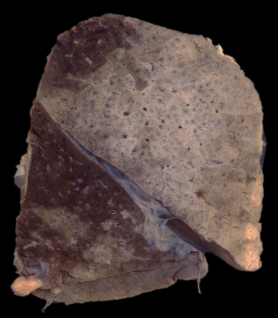
The specimen comprises a sagittal section of left lung, including the left upper lobe and lower lobe. There is a bronchopneumonia characterised by near-confluent consolidation of the left upper lobe (sparing only the apical portion). Small patchy areas of consolidation are also visible in the left lower lobe. An acute fibrinous pleuritis (pleurisy) is present along the visceral pleura in the oblique fissure.
 Right lung
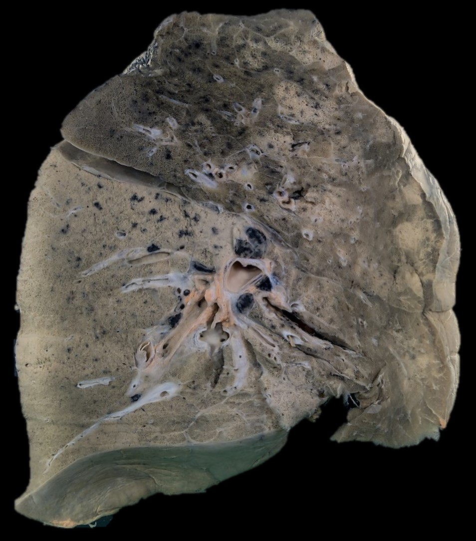
The specimen comprises part of the right lung, and includes the upper, middle and lower lobes. The lower lobe exhibits an almost uniform pale-grey consolidation that closely resembles the appearance of lobar pneumonia in the stage of grey hepatisation. The upper lobe shows multiple large areas of consolidation, which are also beginning to become confluent (bronchopneumonia).
 Left lung
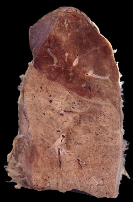
The specimen comprises part of the left lung. The lower lobe is uniformly consolidated (grey hepatisation) by lobar pneumonia. A possible area of consolidation is also discernible in the basal portion of the left upper lobe. There is a conspicuous acute fibrinous pleuritis (pleurisy) along the visceral pleura.
 Right lung
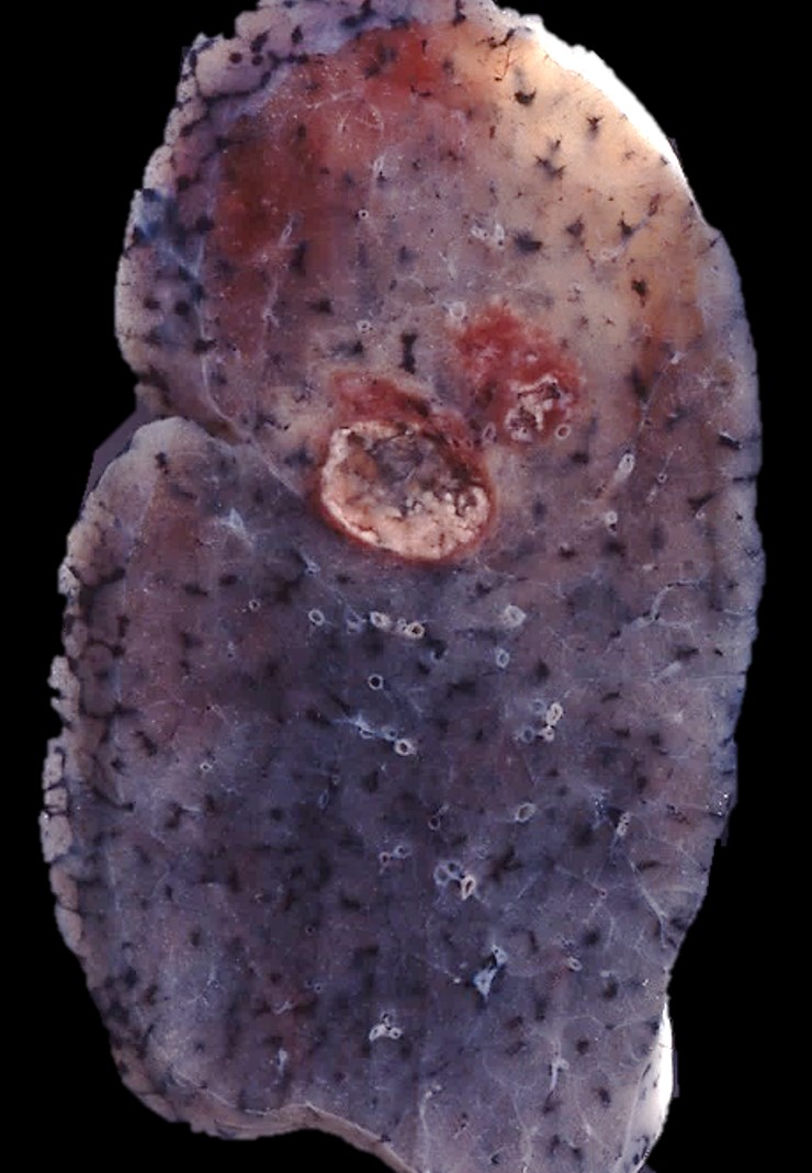
The specimen comprises part of the upper lobe of the right lung, in the centre of which are two lung abscesses, the larger being 2.5cm in diameter. An abscess is a localised collection of pus, in this case in a cavity within the lung parenchyma.


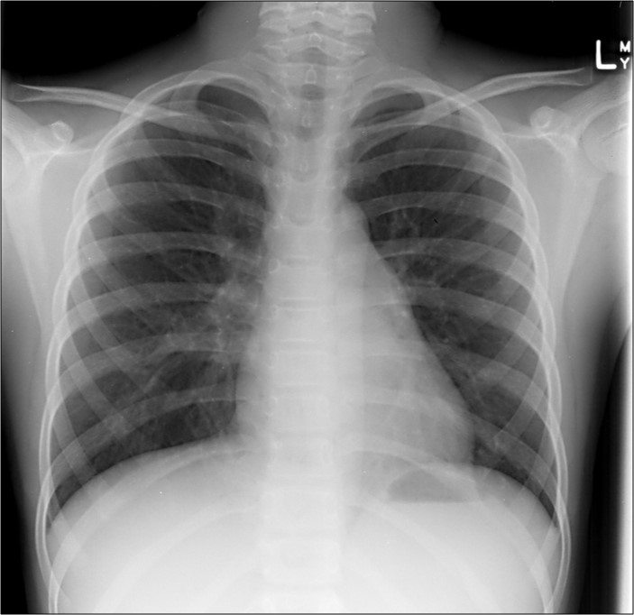
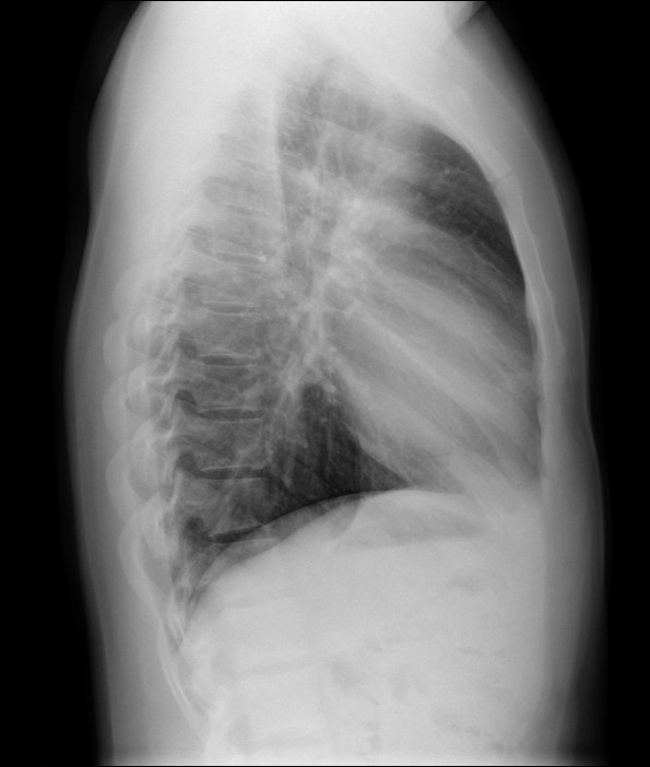
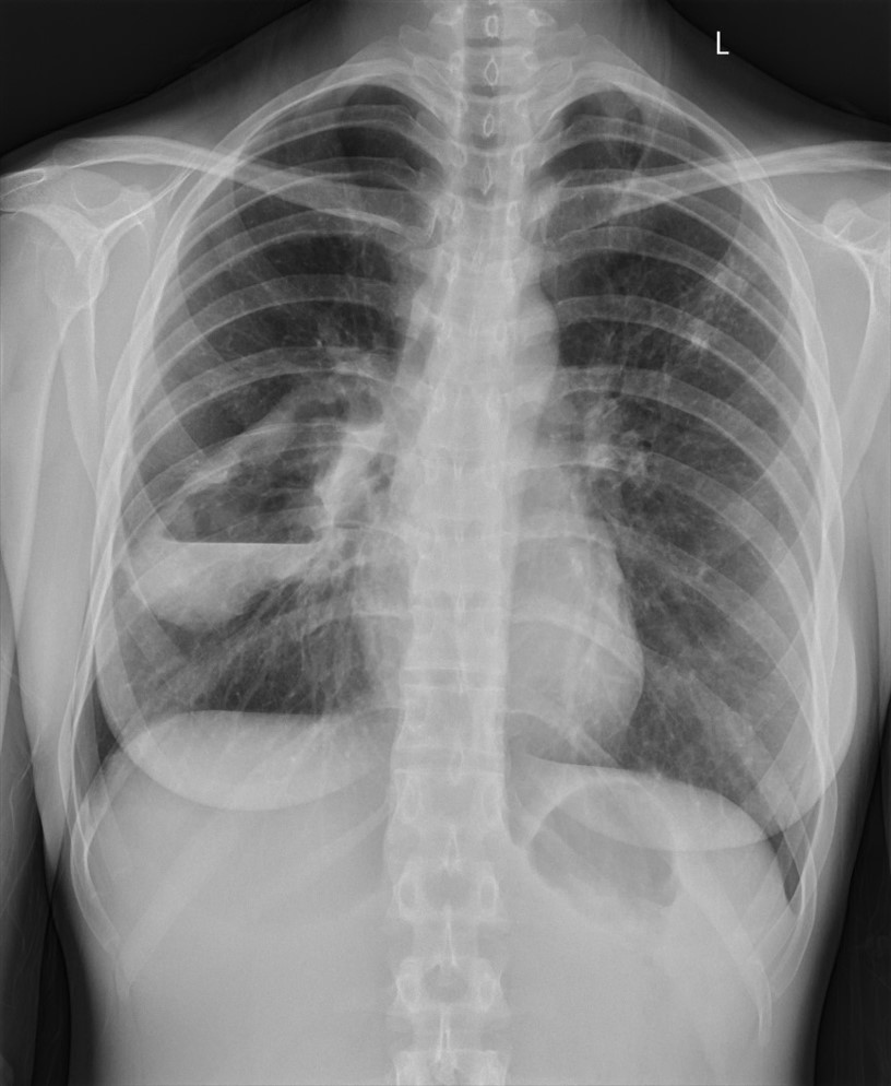
 CXR-1
CXR-1
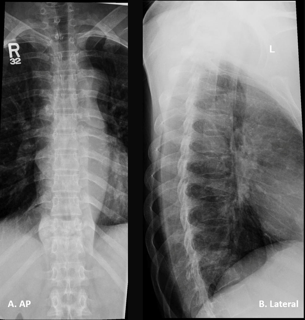
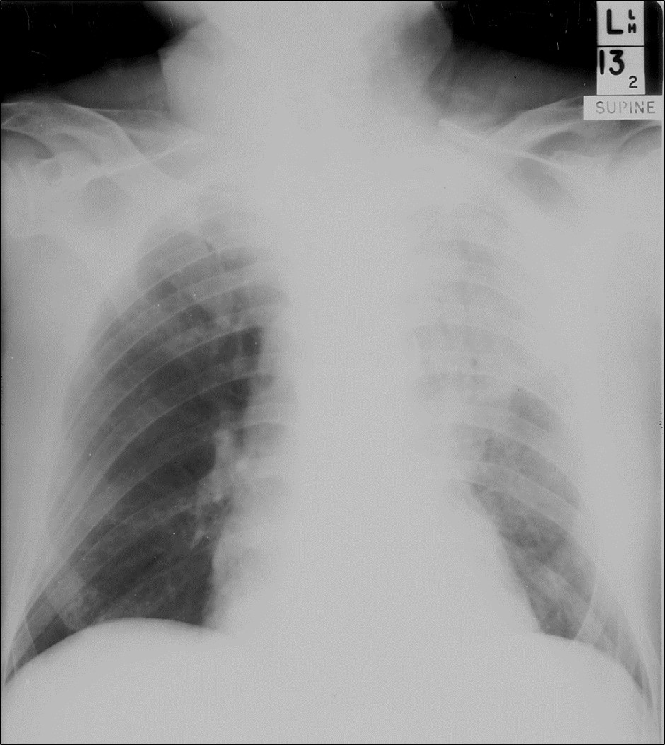
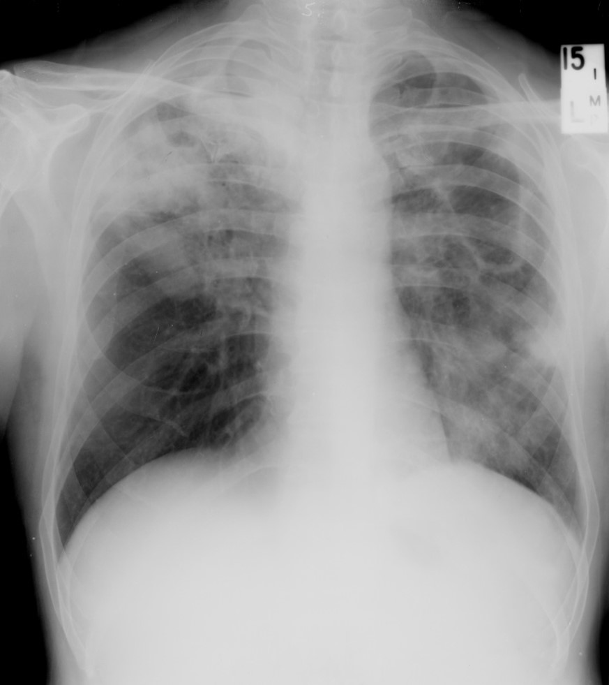
 CXR-2
CXR-2
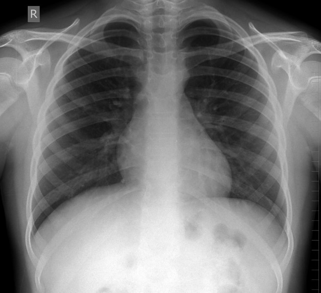
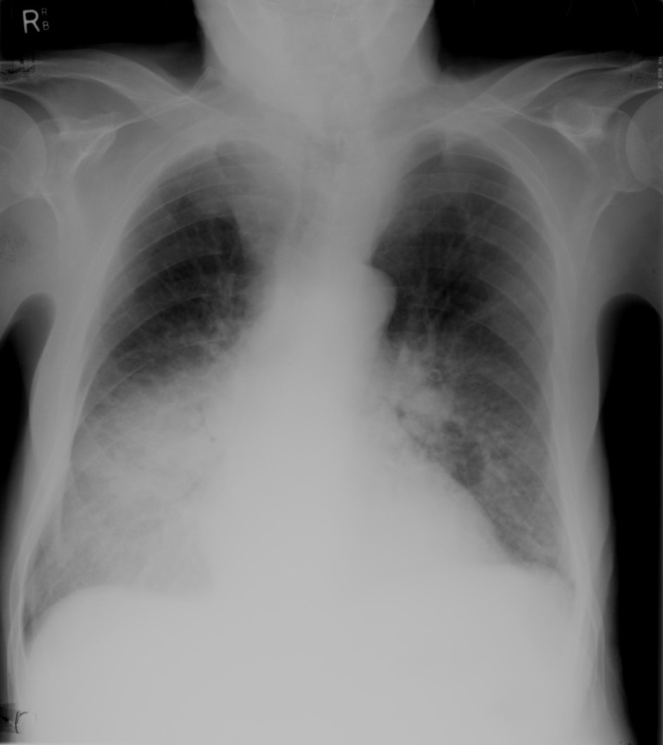
 CXR-3
CXR-3
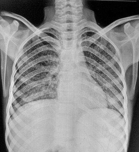
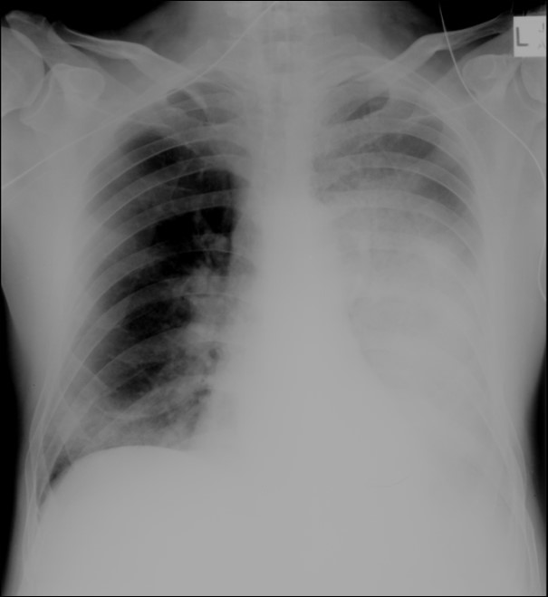
 CXR-4
CXR-4
 XR-4
XR-4